Home

Prononcer ascenseur contourne mitochondria light microscope tondre Ordre alphabétique Énorme

Why can't we see cell organelles such as mitochondria, ribosomes, plastics,  etc under a compound microscope, although it is stained darker than  cytoplasm? - Quora
Why can't we see cell organelles such as mitochondria, ribosomes, plastics, etc under a compound microscope, although it is stained darker than cytoplasm? - Quora

Mitochondria live under Microscope - YouTube

The Shrinking Mitochondrion | The Scientist Magazine®

A new glow for electron microscopy | MIT News | Massachusetts Institute of  Technology
A new glow for electron microscopy | MIT News | Massachusetts Institute of Technology

Mechanisms in Aging Process Illuminated by Light-Powered Mitochondria

Light and electron microscopy of mitochondria in the oocytes of Argulus...  | Download Scientific Diagram
Light and electron microscopy of mitochondria in the oocytes of Argulus... | Download Scientific Diagram

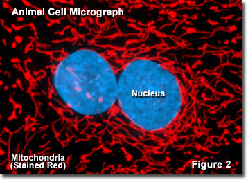
Molecular Expressions Cell Biology: Mitochondria

Mitochondrion - Wikipedia

Are mitochondria the natural cellular light sources?

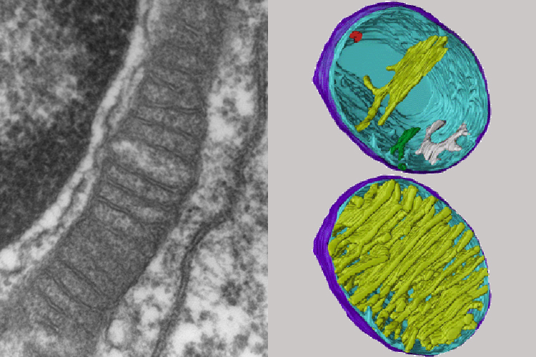
Insight into mitochondrial structure and function from electron tomography  - ScienceDirect
Insight into mitochondrial structure and function from electron tomography - ScienceDirect

Super-resolution microscopy of mitochondria - ScienceDirect

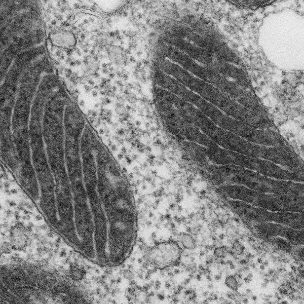
Mitochondria under the microscope — Science Learning Hub

Worms Live Longer with Mitochondria Powered by Light: Preprint | The  Scientist Magazine®
Worms Live Longer with Mitochondria Powered by Light: Preprint | The Scientist Magazine®

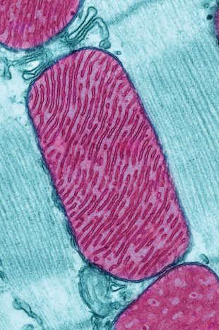
Mitochondrion, TEM - Stock Image - G465/0069 - Science Photo Library

Correlated three-dimensional light and electron microscopy reveals  transformation of mitochondria during apoptosis | Nature Cell Biology
Correlated three-dimensional light and electron microscopy reveals transformation of mitochondria during apoptosis | Nature Cell Biology

Live mitochondria seen in unprecedented detail: photobleaching in STED  microscopy overcome
Live mitochondria seen in unprecedented detail: photobleaching in STED microscopy overcome

Scrutinizing the Microcosm | Natural History Magazine

Mitochondria Under Electron Microscope - YouTube

Mitochondrial swelling detection with a bright-field microscope.... |  Download Scientific Diagram
Mitochondrial swelling detection with a bright-field microscope.... | Download Scientific Diagram

Cell Biology for the Histologist | histology

Ultrastructural examination of ER−mitochondria tethering in HEK 293T... |  Download Scientific Diagram
Ultrastructural examination of ER−mitochondria tethering in HEK 293T... | Download Scientific Diagram

Mitochondria appearance under electron microscope (EM × 6000); A :... |  Download Scientific Diagram
Mitochondria appearance under electron microscope (EM × 6000); A :... | Download Scientific Diagram